Splenic mast cell tumour in a cat
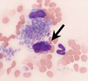
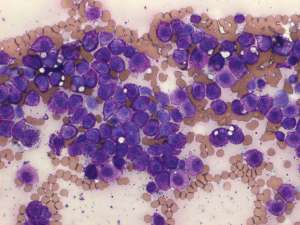
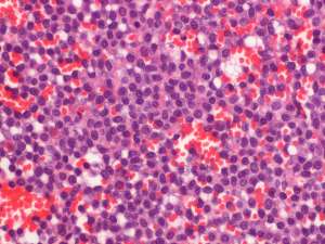
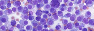
A blood sample was submitted for routine haematology from a 7-year-old female DSH with a history of marked splenomegaly. A fine needle aspirate of the enlarged spleen was performed and subsequently, the spleen was removed and submitted for histopathology.


Final Diagnosis
Splenic mast cell tumour associated with mastocytaemia (systemic mastocytosis)
Discussion
Unlike in dogs, circulating mast cells in cats are not typically associated with non-neoplastic disease and their presence should raise the suspicion of so-called visceral/systemic mastocytosis or a mast cell tumour, particularly splenic. The mastocytaemia in this cat persisted following splenectomy.

