Protozoal dermatitis in a dog
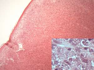
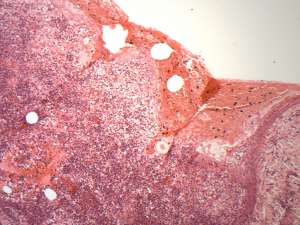
An 8-year-old Boxer presented with chronic variably-sized nodules on the tongue and lips, as well as ulcerated lesions on the skin in multiple locations. The dog was receiving treatment with allopurinol for a previous diagnosis of Leishmania, ciclosporin and amoxycillin-clavulanate, and had previously been treated with prednisolone. The referring vet submitted biopsies of the skin and tongue for histopathology.
Final diagnosis
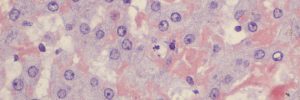
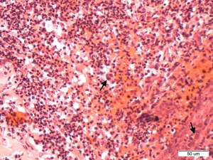
Necrotising dermatitis and granulomatous glossitis with protozoal tachyzoites
Discussion
The morphology of the protozoa is consistent with Toxoplasma sp. or Neospora sp. Given the previous diagnosis of Leishmania in this dog, Leishmania was considered, but the morphology is not consistent with such. As well as other clinical manifestations, both Toxoplasma gondii and Neospora caninum are rarely associated with skin infections in dogs that are immunosuppressed, either naturally or by treatment with immunosuppressive drugs (Hoffmann et al., 2012; Legnani et al., 2016). This dog had received prednisolone and ciclosporin, which may have contributed to immunosuppression and recrudescence of a latent infection. Differentiation between Toxoplasma sp. and Neospora sp. requires PCR and/or immunohistochemistry, which were not performed in this case.
References
Hoffmann et al. (2012). Cutaneous toxoplasmosis in two dogs. J Vet Diag Invest 24(3):636-640.
Legnani et al. (2016). Emergence of cutaneous neosporosis in a dog receiving immunosuppressive therapy: molecular identification and management. Vet Dermatol 27(1)49-e14.