Chordoma in a ferret
HISTORY
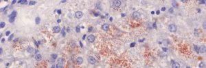
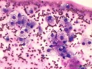
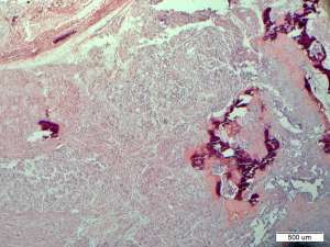
Middle aged to older ferret with large swelling/mass on tail. FNAB was initially performed (see Fig 1) with follow up histopathology (see Fig 2 and 3)
DIAGNOSIS
Chordoma
COMMENT
Chordomas are rare, slow growing neoplasms derived from embryologic remnants of notochord. During foetal development the notochord is replaced by the developing vertebral column, but small remnants remain in the nucleus pulposus of the intervertebral disc. Rests of notochord outside of the intervertebral discs also occur and it is from these which chordomas are thought to develop. While not specific to ferrets, these animals seem to present most commonly with these neoplasms. These masses occur along the axial skeleton but, as in this animal, the distal tail is the most common location for mass development in ferrets. Due to the slow development of the neoplasms early excision is often curative. For those neoplasms along the distal tail complete excision is easily achievable. Masses in other areas along the axial skeleton (e.g. cervical spine) are often less amenable to complete excision and in these cases recurrence is possible. If left unimpeded, metastases can develop in a small subset of cases. In ferrets, this thankfully appears to be a slow process often taking years before clinically apparent metastases are discernible.
References:
Cho et al. Chordoma in the tail of a ferret. Lab Anim Res. 2011.27(1):53-7
Frolich and Donovan. Cervical chordoma in a domestic ferret (Mustela putorius furo) with pulmonary metastasis. JVDI. 2015, Vol. 27(5) 656–659.