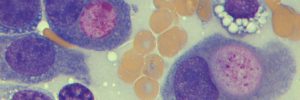
Extratesticular Sertoli cell tumour in a dog
A 10 year old dog presented to the referring vet with a history of a mass in the scrotum. The dog had been castrated 9 years previously. The referring vet excised the mass and submitted it to Cytopath for histopathology.


Final diagnosis
Extratesticular Sertoli cell tumour
Discussion
Sertoli cell tumours are common neoplasms of the testis in dogs, and typically form in entire males of older age. Rarely Sertoli cell tumours may arise in dogs that have been castrated. This phenomenon has also been reported with interstitial/Leydig cell tumours and in neutered male cats (Doxsee et al., 2006). These ‘extratesticular’ tumours may arise in the spermatic cord, in the scrotal skin or at the site of the prescrotal castration incision, and usually many years after the date of castration.
There are multiple possible explanations for the formation of these neoplasms in castrated animals, but the most feasible is neoplastic transformation of cells that have been transplanted inadvertently during previous castration surgery or by trauma. The situations that are considered to predispose to transplantation are incision of the tunica albuginea or embolisation of cells into the veins of the pampiniform plexus caused by excessive pressure placed on the testis or trauma.
Neoplastic transformation of cells within ectopic embryonic testicular remnants or in the case of polyorchidism are considered to be much less likely given the location that the tumours arise in and the rarity of these conditions in dogs and cats.
Therefore, in order to help avoid the formation of extratesticular Sertoli or interstitial/Leydig cell tumours later in life, it is proposed that during castration practitioners avoid exerting excessive pressure on the testis and avoid incising the tunica albuginea.
Reference
Doxsee A, Yager J, Best S & Foster R (2006). Extratesticular interstitial and Sertoli cell tumors in previously neutered dogs and cats: A report of 17 cases. Can Vet J 47:763-766.