Several aspirates were taken from a unilateral submandibular swelling in a 4-year-old, male entire Labrador.
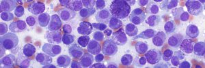
Fig 1. Large clumps of pale blue-grey mucoid material are present, consistent with saliva. This is interspersed with moderate numbers of foamy macrophages.
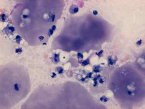
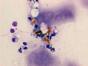
Fig 2. Chronic haemorrhage is a common finding, characterised in this figure by haematoidin (golden yellow pigment) and haemosiderin (green-blue pigment).
Final Diagnosis
Sialocoele
Discussion
This is the typical cell picture associated with a salivary mucocoele (sialocoele).
Their rapid turnaround, cheery disposition and preparedness to discuss cases means I will always choose Cytopath.
Stephen BainesMA VetMB PhD CertVR CertSAS DipECVS DipClinOnc MRCVS, RCVS Specialist in Small Animal Surgery and Oncology, European Specialist in Small Animal SurgeryWillows Veterinary Centre and Referral Service, Shirley, West Midlands