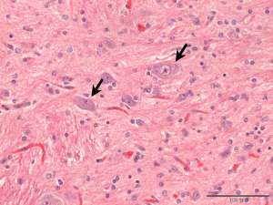
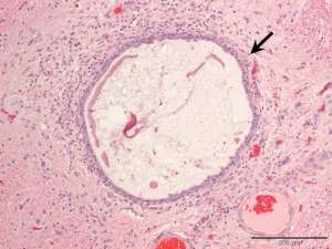
These are histopathology sections from an ovarian mass discovered at the time of spay of a 1-year-old Labrador Retriever.
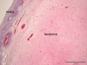
Fig 1. The bulk of the ovarian mass in this section is composed of disorganised neural tissue in which numerous glial cells and neurons are scattered, resembling disorganised brain/spinal cord.
Fig 2. Arrows indicate the neurons.
Fig 3. Scattered within the mass are small cystic structures (arrow) that are lined by uniform cuboidal to columnar epithelial cells that occasionally have apical cilia. The cysts contain small amounts of eosinophilic/proteinaceous material.
Final Diagnosis
Ovarian teratoma
Discussion
Ovarian teratomas are uncommon primary ovarian tumours that are derived from two or more germinal cell layers; in this case a neural and epithelial component are identified. Most teratomas are benign and malignant forms are rare.
I have been using the services of Cytopath for the last 10 years and wouldn’t hesitate to recommend their detailed and efficient reporting. They are always helpful, friendly and happy to discuss cases. Their speed of reporting urgent results is second to none and they never fail to deliver. Thank you team Cytopath for providing such a top notch service to us!
Kirsty RoeBVSc CertSAM DipACVIM DipECVIM-CA MRCVSWillows Veterinary Centre and Referral Service, Shirley, West Midlands