Mycobacterial infection in an eye
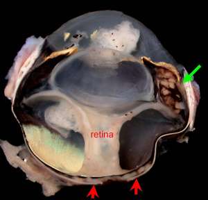
Enucleation was performed in a 2-year-old male DSH cat that presented with a florid uveitis in the left eye. The eye was blind and retinal detachment was confirmed on ocular ultrasound
Final Diagnosis
Pyogranulomatous uveitis and retinal detachment associated with mycobacterial infection
Discussion
Mycobacterial infection is an important differential in the case of pyogranulomatous uveitis. Many of these cats will present with a predominant posterior uveitis/choroiditis and associated retinal detachment is common. At the time of presentation, cats with ocular disease may or may not have concurrent signs of systemic disease (e.g. pyogranulomatous to granulomatous panniculitis, pneumonia, arthritis/synovitis or nephritis).
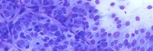
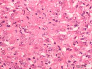
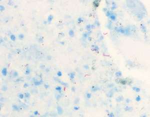
Mycobacteria are typically not evident on routine H&E-stained sections and special stains (e.g. Ziehl-Neelson) are required for their detection; in addition, the actual number of organisms is frequently very low and a high index of suspicion is helpful to confirm the diagnosis. In order to speciate further, culture of fresh tissue remains the gold standard. PCR can also be performed (on fresh or formalin-fixed tissue) in an attempt to detect the presence of the M.tuberculosis complex (M. tuberculosis, M.bovis and M.microti) or to detect non-tuberculosis mycobacteria (e.g. M.avium, M.fortuitum), however false negatives do occur.