Lingual Granular Cell tumour in a dog
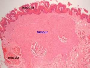
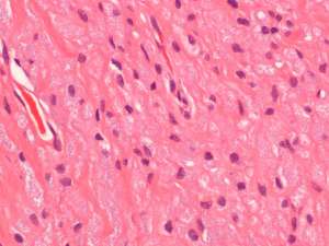
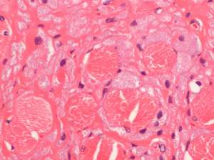
These are sections taken from a mass in the tongue of 10-year-old male Labrador.


Final Diagnosis
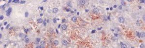
Granular cell tumour
Discussion
These tumours tend to be slowly progressive and are believed to be neural in origin. In dogs, the tongue is the most commonly affected site and so this is an important differential diagnosis for a tumour affecting the tongue. Currently, the rate of recurrence following complete excision and the risk of metastasis are believed to be low.

