Corneal inclusion cysts in a dog
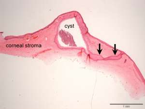
The following are H&E sections prepared from a keratectomy specimen in a 4-year-old male neutered pug. The pug had a history of a deep corneal ulcer in the right eye which was surgically repaired using a conjunctival pedicle graft. Six months later, the superficial cornea had developed two discrete, smooth, creamy yellow-white nodular lesions (up to 2.0 x 1.5 x 3.0mm in diameter) at the site of the conjunctival graft. These lesions were clinically consistent with corneal inclusion cysts and a keratectomy was performed to excise them.
Final Diagnosis
Multiple corneal inclusion cysts
Discussion
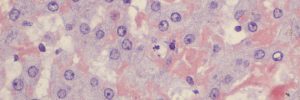
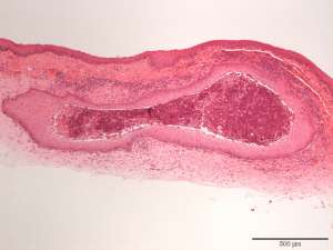
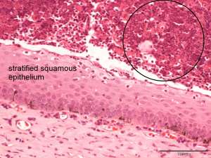
Corneal inclusion cysts are uncommon, benign lesions in domestic animals. They may be congenital however the vast majority are acquired and are usually associated with previous trauma, including surgical trauma (e.g. at the site of sutures). The most likely pathogenesis is a downgrowth or embedding of corneal epithelium within the corneal stroma with associated epithelial proliferation and cyst formation. The cysts typically contain abundant neutrophils and squames which is what will be seen cytologically if these lesions are aspirated. Many of the cysts tend to be relatively superficial in the corneal stroma and can be excised by keratectomy.