Fungal keratitis in a dog
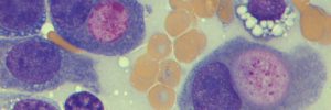
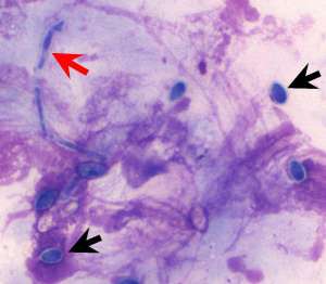
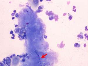
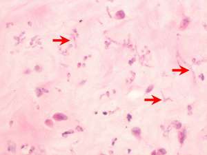
Corneal scrapes, a keratectomy sample and swab were submitted from a 1-year-old Border Collie, taken from a painful white corneal opacity.
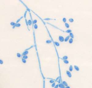
Final Diagnosis
Fungal keratitis caused by Scedosporium sp.
Discussion
The fungus was identified as Scedosporium prolificans. This is an earth-borne opportunistic fungus that is isolated in soil and water. It is an emerging cause of fungal infections and its identification is important because it is often resistent to antifungal therapy hence necessitating surgical intervention. Fungal keratitis is not uncommon in horses and human patients but is not a common diagnosis in dogs. Corneal injury and immunocompromise (e.g. secondary to long term corticosteroid use) likely predispose to the development of fungal keratitis but a history of corneal trauma/immunosuppression is not always present in reported cases. Currently, cases of Scedosporium sp keratitis have been reported in humans, chickens and dogs.