An enlarged sublumbar lymph node in a hypercalcaemic dog
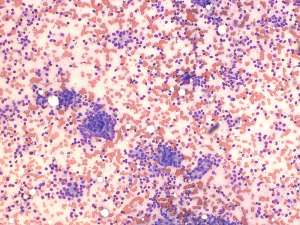
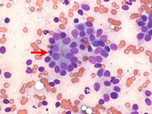
These are fine needle aspirates taken from an enlarged left sublumbar lymph node in a 10-year-old female Labrador. There is a clinical history of hypercalcaemia and no other lymph nodes are reported to be enlarged.
Final Diagnosis
Metastatic carcinoma
Discussion
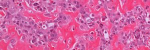
The presence of lymph node enlargement and hypercalcaemia is not necessarily indicative of lymphoma and the epithelial cell population present in this enlarged sublumbar lymph node is most reminiscent of that seen in anal sac gland adenocarcinomas. Anal sac gland adenocarcinomas are malignant neoplasms and the epithelial cells appear deceptively bland on cytology. These tumours commonly metastasise to the regional lymph nodes, particularly the sublumbar and iliac lymph nodes and a proportion of dogs may develop paraneoplastic hypercalcaemia.