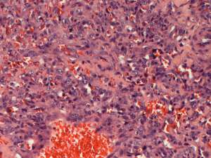
Cutaneous haemangiosarcoma in an elderly dog
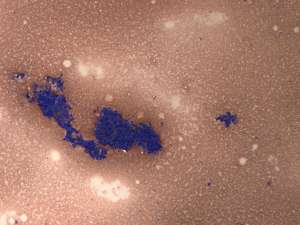
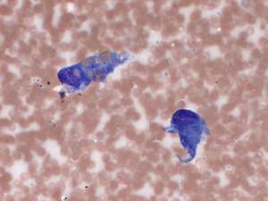
An 11-year-old Lurcher presented with a solitary subcutaneous mass on its left flank. Fine needle aspirates were initally taken from the mass and following the cytology result, surgical resection and histopathology were performed.
Final Diagnosis
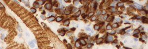
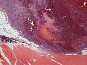
Haemangiosarcoma
Discussion
Haemangiosarcomas are malignant, locally infiltrative neoplasms of vascular (endothelial) origin and post-surgical recurrence can be problematic. These tumours can potentially arise within any tissue, including the skin, deep subcutis and/or muscle. Primary cutaneous tumours do carry metastatic potential (especially to lungs) but the metastatic rate is lower compared to their visceral counterparts. Less commonly, metastatic lesions may occur in the skin from a primary haemangiosarcoma elsewhere (e.g. splenic, hepatic, cardiac or bone).