Malignant transformation of a viral papilloma in a dog
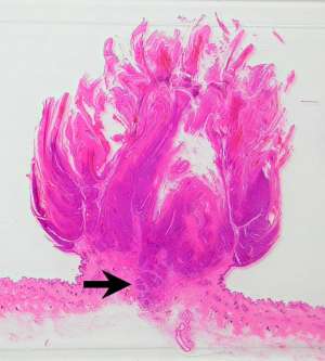
The following are histopathology (H&E) sections of a solitary, exophytic papilliform skin mass that was surgically resected from the dorsum of a 1-year-old male Labrador Retriever.
Final Diagnosis
Squamous cell carcinoma (ex-viral papilloma)
Discussion
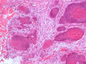
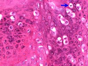
Cutaneous viral papillomas in dogs usually occur in dogs under two years of age and are caused by a papillomavirus. The majority of these tumours undergo regression over a course of weeks to months. In rare cases, malignant transformation to a squamous cell carcinoma can occur. In this case, although the clinical appearance was typical of a benign papilloma, the presence of invasive nests of squamous cells at the base of the tumour indicates malignant transformation. From a practical point of view, biopsy specimens of papillomatous lesions should therefore ideally include a deep margin so that this can be properly examined for any evidence of invasive behaviour/malignancy.