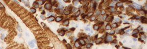
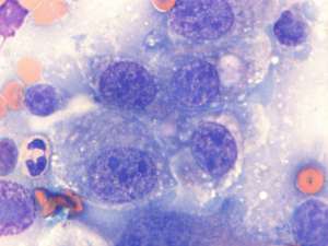
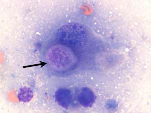
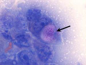
Transitional cell carcinoma (TCC) in a dog
The following are cytology smears prepared from a urethral catheter suction biopsy of a urinary bladder mass in a 12-year-old female Labrador Retriever with a clinical history of dysuria and haematuria.
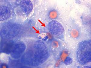
Final Diagnosis
Carcinoma, consistent with a transitional cell carcinoma (TCC)
Discussion
Transitional cell carcinomas are malignant and locally invasive neoplasms. They most commonly involve the urinary bladder, urethra, prostate (secondary to prostatic invasion by a prostatic urethral TCC) and kidney. Unfortunately, these tumours carry a high risk of metastasis, particularly to the regional sublumbar lymph nodes, lungs and bone (e.g. pelvic and lumbar).