Signet-ring cell carcinoma in the stomach of a dog
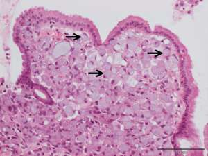
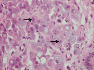
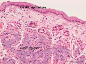
The following are histopathology (H&E) sections from endoscopic biopsies taken from an area of diffuse thickening in the lesser curvature of the stomach of a 10-year-old male Patterdale Terrier. Clinical signs included chronic vomiting and weight loss.
Final Diagnosis
Signet-ring cell gastric carcinoma
Discussion
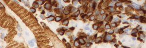
Signet-ring cell carcinomas are a variant of adenocarcinoma in which the cytoplasm of the neoplastic epithelial cells distends with mucin causing peripheralisation of the nucleus and resulting in the morphological appearance of a ‘signet ring’. These signet-ring cells are typically round to polygonal and do not form typical glandular structures. As with all variants of gastric adenocarcinomas, dogs commonly present with advanced disease and local invasion, ulceration, fibrosis and distant metastasis is common. Metastasis most commonly occurs to the regional gastric and gastroduodenal lymph nodes, omentum and diaphragm and less commonly to the lungs, liver and spleen.