Lungworm infection associated with hypercalcaemia in a dog
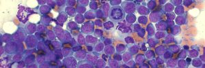
A bronchoalveolar lavage (BAL) was performed in a 4-year-old crossbreed dog with a clinical history of coughing. Air-dried direct smears were made at the time of sampling and were submitted together with the EDTA fluid sample for cytological evaluation. Thoracic radiographs revealed an interstitial lung pattern and the dog was also mildly hypercalcaemic (both total and ionised calcium were increased).
Final Diagnosis
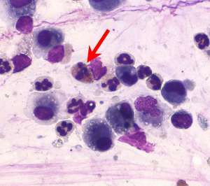
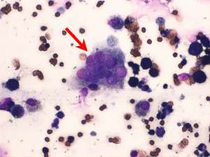
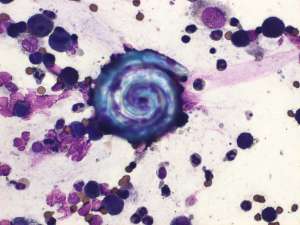
Lungworm (Angiostrongylus vasorum) infection associated with pyogranulomatous pneumonia and hypercalcaemia
Discussion
Hypercalcaemia associated with lungworm infection is not common but remains an important differential to consider in hypercalcaemic dogs. Hypercalcaemia secondary to granulomatous inflammation is well-recognised and is likely due to the dysregulation of calcitriol production by activated macrophages. BALs from dogs infected with Angiostrongylus vasorum do not necessarily reflect eosinophilic inflammation and some cases are associated with a pyogranulomatous to granulomatous response with very few or no eosinophils. The calcium levels in this dog returned to normal following treatment for lungworm.