
Cutaneous toxoplasmosis in a cat
Fine needle aspirates were taken from a single, 3 x 1.5cm cutaneous mass on the neck (in the region of the thyroid gland) of a 14-year-old, female neutered DSH. The cat had a history of chronic weight loss. FeLV/FIV status was negative.
Discussion
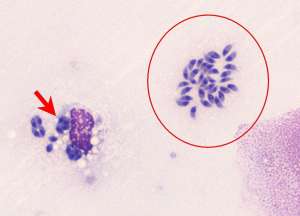
This cell harvest is indicative of a protozoal infection. The two main differentials for the organism include Toxoplasma gondii and Neospora caninum with the former being more likely given the affected species and similarities to previously reported cases of cutaneous toxoplasmosis in cats. Differentiation between Toxoplasma sp and Neospora sp typically requires immunohistochemistry, electron microscopy or PCR, with PCR probably being the most useful.
Cutaneous toxoplasmosis in cats is uncommon but it is typically a manifestation of systemic infection. Cutaneous lesions are usually nodular with or without ulceration, may be solitary or multiple and usually reflect dermatitis and/or necrotising panniculitis and vasculitis. Systemic toxoplasmosis most commonly results in a necrotising interstitial pneumonia, hepatitis, lymphadenitis and myocarditis. Affected cats often present with respiratory distress due to the interstitial pneumonia and pleural effusion but this may be absent in cats admitted for preliminary cutaneous lesions. Underlying disease may be associated with toxoplasmosis and typical examples include immunosuppression by glucocorticoids, FIP, FIV and FeLV. An underlying cause was not identified in this cat.