An iridociliary adenoma in a Labrador Retriever
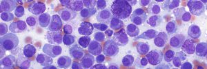
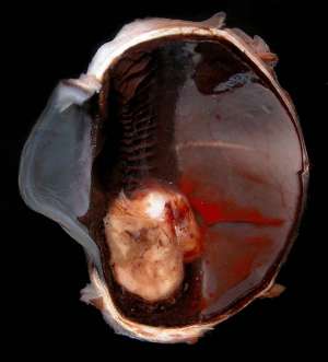
The following are pictures of the gross and histopathological specimens taken from the enucleated left eye of a 12-year-old male Labrador Retriever that presented with chronic intraocular haemorrhage and glaucoma.

Final Diagnosis
Iridociliary adenoma
Discussion
Iridociliary adenomas are the second most common primary intraocular tumours in dogs after anterior uveal melanomas. They arise most commonly from the non-pigmented or pigmented ciliary body epithelium and less commonly from the posterior iris epithelium. Frequent complications associated with the tumour include intraocular haemorrhage, anterior uveitis and secondary glaucoma. Enucleation is curative. Iridociliary adenomas can also occur in cats, however they are less common in this species