
A cutaneous infiltrative lipoma in a 6-year-old female Labrador Retriever
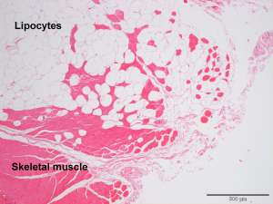
The following are H&E sections from an incisional biopsy taken from a subcuticular mass of the left flank that clinically appears firmly fixed to the underlying tissue.
Final Diagnosis
Infiltrative lipoma
Discussion
These are uncommon neoplasms in the dog and are typically nonencapsulated, infiltrate along fascial planes and invade muscle. Although they do not undergo malignant transformation or possess any metastatic potential, they may require more radical surgical excision due to their infiltrative nature. Females are predisposed and commonly affected sites include the neck, trunk and proximal limbs.
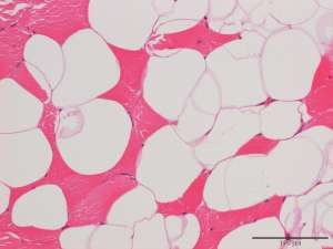
The neoplastic lipocytes of an infiltrative lipoma essentially resemble the lipocytes of normal adipose tissue and confirmation of the diagnosis requires histopathological evaluation for infiltrative behaviour. It is worth noting that obese dogs may have a significant amount of fat within underlying skeletal muscle bundles and therefore small incisional or trucut biopsies may not be sufficient to support a suspected diagnosis of infiltrative lipoma.