FIP in a cat - an uncommon presentation
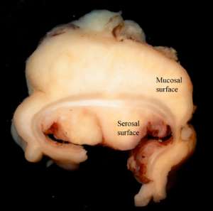
An 8-month-old DSH kitten with a history of chronic diarrhoea and vomiting presented with a palpable colonic mass that was surgically resected and submitted for histopathological evaluation. No other lesions were grossly evident at the time of exploratory laparotomy.
Final diagnosis
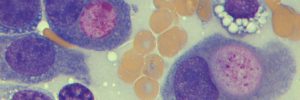
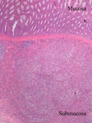
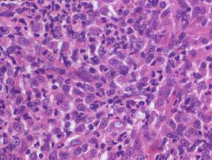
Feline infectious peritonitis (FIP) associated with focal nodular pyogranulomatous colitis
Discussion
This case is an example of an uncommon but recognised intestinal presentation of a common infectious disease. FIP is an invariably fatal and sporadic viral disease of domestic and wild felids caused by feline corona virus (FCoV).
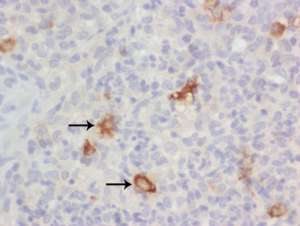
The majority of cats with non-effusive (dry) FIP present with pyogranulomatous inflammation or granulomas within the brain, kidneys, eye, lymph nodes and/or omentum. Occasionally however, cats affected with noneffusive (dry) FIP may present with chronic vomiting and diarrhoea associated with a palpable mass at the ileocecocolic junction or adjacent colon. On gross examination, the appearance of the mass can resemble that of lymphoma but FIP should remain an important differential diagnosis, particularly in cats of appropriate signalment.