
Scleral rupture in a cat
A 5-year-old, male cat was involved in a road traffic accident (RTA) and suffered proptosis of the right globe, which was subsequently repositioned. One month later the eye was enucleated due to persistent uveitis, secondary glaucoma and blindness.
Gross Findings
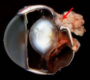
The main lesion (see figure below) is a focal rupture of the posterior sclera. Note the adherent extraocular fat and muscle at the rupture site (arrow). The lens is displaced posteriorly. The retina is detached and coats the posterior lens surface admixed with chronic vitreal haemorrhage.
Final Diagnosis
Traumatic posterior scleral rupture
Discussion
Scleral rupture occurs most commonly as a result of blunt trauma, for which RTAs are the leading cause in cats. Because the majority of ruptures are located within the posterior sclera, they are often not identified on initial clinical examination but can usually be confirmed using ultrasound.
The acute decompressive forces associated with blunt trauma to the globe that result in scleral rupture are often accompanied by significant injury to the internal ocular structures subsequently resulting in permanent blindness. Associated dislocation of the lens secondary to severe trauma is not uncommon such as in this case and in some cases, the lens can rupture or actually be extruded through the scleral wound.
If enucleation is not performed, eventual shrinkage of the globe will occur resulting in phthisis bulbi.