Neoplastic pleural effusion in a dog
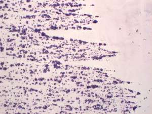
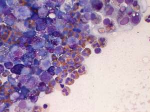
Samples of pleural fluid were submitted from a 10-year-old Golden Retriever for cytological evaluation.
Final Diagnosis
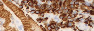
Mast cell neoplasia
Discussion
The dog had a history of multiple cutaneous mast cell tumours and in the absence of other primary lesions, the neoplastic effusion is consistent with metastatic disease. Similar to carcinomatosis, regardless of the site of primary neoplasia, the neoplastic cells can exude via the lymphatics into the thoracic, pericardial and abdominal cavities, resulting in a neoplastic exudate.