Extraskeletal osteosarcoma in a dog
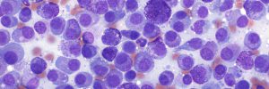
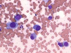
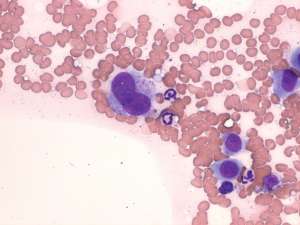
A 1-year-old male neutered Dalmatian presented with a firm 4x3x3cm subcuticular mass overlying the right chest wall. There was no clinical or radiological association of the mass with the underlying ribs and no other lesions were found. A fine needle aspirate was performed and following the results, the mass was surgically resected and submitted for histopathology.
Final Diagnosis
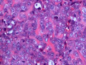
Extraskeletal osteosarcoma
Discussion
The presence of a solitary subcuticular neoplasm in the absence of primary bone involvement is typical of an extraskeletal osteosarcoma. The majority of reported extraskeletal osteosarcomas are in the mammary gland and other sites less frequently involved include the subcutaneous tissue, liver, spleen, thyroid gland, intestinal tract, eye and muscle. Oesophageal osteosarcomas in association with Spirocerca lupi infection in dogs is a well-recognised phenomenon. Unfortunately, similar to primary bone osteosarcomas, the risk of metastasis is high.