Ductal plate malformation
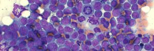
This is a 5-month-old German Shepherd dog that was being worked up for elevated liver enzymes on routine blood work. Ultrasound examination revealed an extrahepatic shunt.
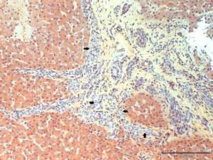
Final Diagnosis
Moderate, multifocal, bridging portal fibrosis with moderate to marked bile ductule proliferation (ductal plate malformation)
Discussion
During early embryonic development of the liver there is a plate of bi-potential hepatoblasts (ductal or limiting plate) that surround mesenchyme and the embryonic portal vein which will later form the portal triads. The hepatoblasts represent a common progenitor population from which the biliary epithelium and hepatocytes will develop. Malformations in the ductal plate are rare in dogs and develop as a result of a lack of remodelling of the embryonic biliary system leading to retention of rudimentary bile ductules. The retained fibrous tissue associated with the embryonic biliary system will impede normal blood flow in affected livers causing hypertension, which in its own right can cause vascular shunts. As the vascular system develops somewhat synchronously with the ductal plate, concurrent vascular anomalies (e.g. shunts, portal hypoplasia) can occur. Vascular changes consistent with portal hypertension are also observed in this liver.