An orbital meningioma in a 10-year-old female Bull Mastiff
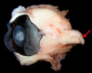
The following are pictures of the gross and histopathological specimens taken from the exenterated orbit of 10-year-old female Bull Mastiff that presented with exophthalmos, protrusion of the third eyelid and blindness in the left eye. Ophthalmic evaluation revealed a swollen optic nerve head and indentation of the posterior sclera. An MRI scan confirmed the presence of an orbital mass and orbital exenteration was performed via an orbitotomy.
Final Diagnosis
Canine orbital meningioma
Discussion
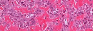
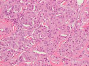
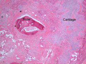
This is a slow-growing neoplasm that arises from the extradural arachnoid cap cells and typically surrounds the optic nerve to form a cone-shaped orbital mass. The neoplastic cells infiltrate the surrounding orbital fat, skeletal muscle and, in some cases, the posterior globe itself. Complete resection can be challenging and any remaining neoplastic tissue will result in local recurrence and/or extension into the optic foramen, optic chiasm or brain. Metastasis is rare. The histopathological pattern is meningotheliomatous whereby neoplastic cells are most often epithelial-like in appearance and hence the tumour must not be confused with a carcinoma. An additional common finding in these tumours is osseous and chondroid metaplasia, a feature that can be diagnostically useful.