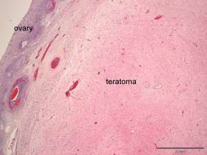
Ovarian teratoma

These are histopathology sections from an ovarian mass discovered at the time of spay of a 1-year-old Labrador Retriever.

Fig 1. The bulk of the ovarian mass in this section is composed of disorganised neural tissue in which numerous glial cells and neurons are scattered, resembling disorganised brain/spinal cord.
Fig 2. Arrows indicate the neurons.
Fig 3. Scattered within the mass are small cystic structures (arrow) that are lined by uniform cuboidal to columnar epithelial cells that occasionally have apical cilia. The cysts contain small amounts of eosinophilic/proteinaceous material.

Final Diagnosis

Ovarian teratoma

Discussion

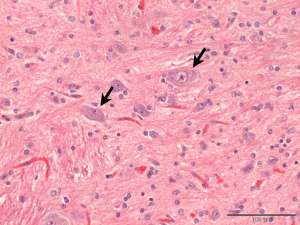
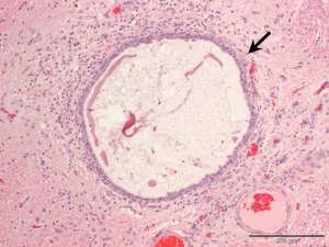
Ovarian teratomas are uncommon primary ovarian tumours that are derived from two or more germinal cell layers; in this case a neural and epithelial component are identified. Most teratomas are benign and malignant forms are rare.