Glomerulonephritis in a dog
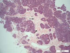
A 9-year-old Yorkshire terrier presented for acute haemorrhagic gastroenteritis that resolved on symptomatic treatment. A faecal culture at the time was positive for Campylobacter sp. Two weeks later, the dog re-presented with ascites and subcuticular oedema. Biochemical findings included a significant hypoalbuminaemia (7g/dL), hypoglobulinaemia, hypocholesterolaemia and azotaemia. The urine SG was 1.014. The urine protein: creatinine ratio was 52 (normal is less than 0.5). A renal biopsy was taken and the figures below focus on the glomerular lesions present.
Final Diagnosis
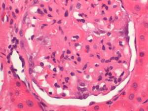
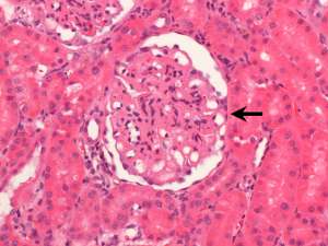
Diffuse generalised membranoproliferative glomerulonephritis
Discussion
The membranoproliferative glomerulonephritis in this dog accounts for the marked proteinuria and azotaemia. It is not aetiologically specific however such lesions typically reflect the deposition of immune-complexes or activation of complement within glomeruli. The initiating cause in this case is speculative but given the clinical history and sequence of events, a postinfectious glomerulonephritis is suspected. Interestingly, in human patients, acute postinfectious glomerulonephritis associated with Campylobacter jejuni enteritis has been published and this organism's potential to trigger immune-mediated renal disease is recognised.