Cutaneous angiomatosis in a dog
The following are histopathology (H&E) sections of a solitary, ulcerated cutaneous mass surgically resected from the neck of a 1-year-old male Labrador Retriever.
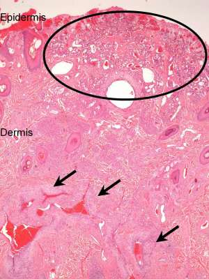
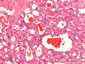
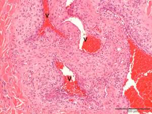
Final Diagnosis
Cutaneous angiomatosis
Discussion
Angiomatosis is an umbrella term used to denote various vasoproliferative lesions including vascular malformations and hyperplasia. The majority of dogs affected are young although lesions can also occur in adult/older dogs. Despite the bland cellular appearance of these lesions, they can be locally infiltrative and progressive. Although any site can be affected, lesions most commonly occur on the feet and are often associated with bony destruction. Metastasis has not been reported however incomplete excision will result in local recurrence and additional lesions can occur.