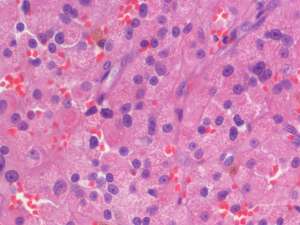
Leydig cell tumour
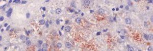
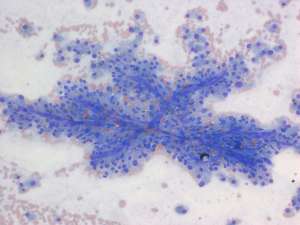
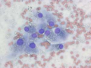
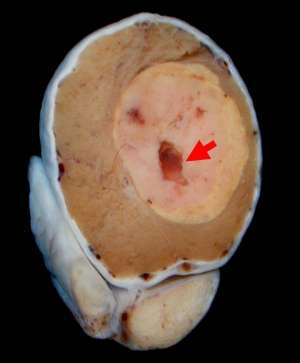
A non-painful mass was detected in the left testicle of a 12-year-old male Labrador during a routine examination at the time of vaccination. The mass was aspirated and following the cytology results, castration was performed and the affected testicle submitted for histopathology.
Final Diagnosis
Leydig (interstitial) cell tumour
Discussion
Leydig cell tumours are common tumours of the testis and may be solitary or multiple. These tumours arise from the interstitial cells of the testis which under normal conditions are responsible for androgen production. The vast majority of Leydig cell tumours are behaviourally benign and metastatic spread is exceptionally rare. Occasionally Leydig cell tumours may be hormonally active which can result in concurrent prostatic and/or perianal gland hyperplasia. These changes usually regress following castration