Laryngeal rhabdomyoma in a dog
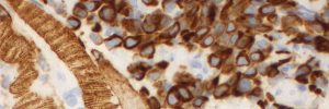
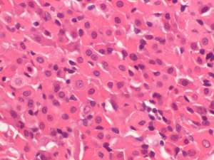
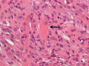
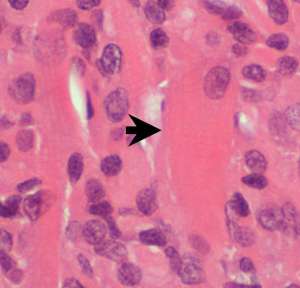
Biopsies taken from a mass protruding into the lumen of the larynx in a 12-year-old crossbreed dog with a history of increased upper respiratory noise.
Final Diagnosis
Laryngeal rhabdomyoma
Discussion
These are uncommon tumours of skeletal muscle but when they occur in the dog, they most commonly occur in the larynx. They typically protrude into the larynx and some may display low grade locally infiltrative behaviour. The presence of well-differentiated neoplastic cells, limited invasiveness and a low mitotic rate support a benign tumour and the low number of reported cases suggest a benign clinical course with regards to local recurrence and metastatic disease.