Uveodermatological syndrome in an Akita
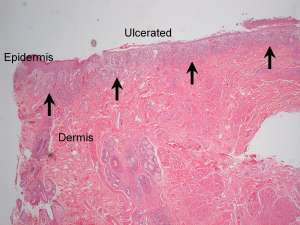
The following are histopathology (H&E) sections of depigmented and erythematous skin from the dorsal muzzle of a 2-year-old male Akita. The dog also has concurrent bilateral uveitis.
Final Diagnosis
Granulomatous lichenoid dermatitis
Discussion
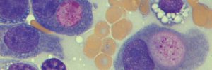
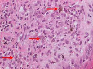
The histopathological lesion of a granulomatous lichenoid (band-like) dermatitis associated with the accumulation of fine dust-like melanin pigment within macrophages, together with the breed and clinical findings, is indicative of uveodermatological syndrome. This is also known as Vogt-Koyanagi-Harada-like syndrome (VKH).
This is an autoimmune disease and the proposed mechanism is a cell-mediated hypersensitivity reaction against melanin and related proteins. In dogs, the uveal tract and skin are typically affected resulting in a granulomatous panuveitis and dermatitis. Ocular signs often preceed the cutaneous lesions and prompt recognition and treatment of this disease is important to prevent permanent blindness. The disease can affect multiple breeds, however Akitas, Samoyeds, Siberian Huskies, Alaskan Malamutes and Chow Chows are particularly predisposed. Cutaneous lesions (which resemble those of other auto-immune diseases) include symmetrical facial depigmentation, erythema and crusting. Lesions particularly affect the periorbital skin, dorsal muzzle, nasal planum and lips although the scrotum, vulva and pawpads can also be affected. Biopsies should be taken from regions of depigmentation, erythema and crusting – clearly ulcerated and infected areas are best avoided.