Urticarial allergic reaction in a dog
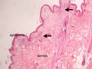
This is a skin biopsy from a 3-year-old Jack Russell Terrier. The dog developed a generalised erythematous rash and limb oedema within 36 hours of antibiotic and anti-inflammatory treatment for a bout of vomiting and diarrhoea.
Final Diagnosis
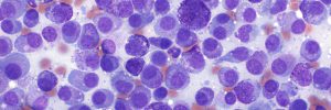
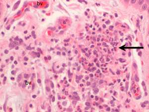
Superficial perivascular to interstitial eosinophilic dermatitis
Discussion
The histopathology together with the clinical history is consistent with urticarial allergic eruption, associated with a hypersensitivity reaction. It is not cause-specific but an adverse drug reaction is one of the most commonly reported causes and was suspected in this particular case. Other causes include insect/arthropod bites, vaccines and food allergy.