
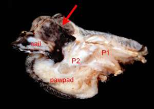
Subungual melanoma in a dog
An 8-year-old male Rottweiler presented with swollen digit on the left frontleg and radiographs indicated lysis of the third phalanx (P3). Amputation of the digit was performed and submitted for histopathological evaluation.
Final Diagnosis
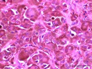
Subungual malignant melanoma
Discussion
Subungual melanomas arise from the melanocytes of the nailbed. These tumours are usually slowly progressive and are commonly associated with invasion and lysis of P3 at the time of diagnosis. Breed predispositions include the Scottish terrier, Schnauzer, Rottweiler, Golden retriever and Irish setter. The major clinical differential is a squamous cell carcinoma which is the most common malignancy affecting the canine digit. Reported metastatic rates of subungual melanomas are reportedly higher than other cutaneous melanomas ranging from 38-58% with a 1-year survival rate of 42-70%. Another recent study reported a metastatic rate of 50% with a mean disease-free interval of 322 days.