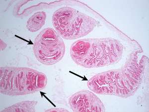
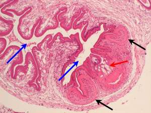
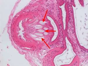
Subcuticular coenurus in a rabbit
The following are H&E sections of a discrete subcuticular cyst associated with the underlying skeletal muscles in the shoulder region of a 2-year-old male rabbit. The cyst was surgically removed intact and submitted for histopathological examination.
Final Diagnosis
Subcuticular coenurus
Discussion
This is essentially a cystic structure that contains cestode (tapeworm) larvae and surgical excision from this location is the treatment of choice. Given the species and tissues involved, this is consistent with a coenurus of Taenia serialis in which the intermediate host is the rabbit or hare and the definitive host is the dog or fox. Fluid-filled cysts typically develop under the skin, between muscles and within the fascia. Retrobulbar involvement has also been reported. Infection in wild rabbits and hares is more common, however pet rabbits typically become infected when their bedding or food is contaminated with canid faeces.