Sebaceous adenitis in a dog
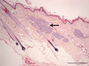
Skin biopsies have been taken from areas of scaling and crusting on the dorsal neck and trunk regions of a 2-year-old female poodle.
Final Diagnosis
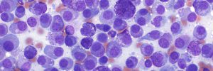
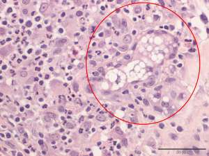
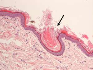
Granulomatous sebaceous adenitis associated with sebaceous gland loss and hyperkeratosis
Discussion
Sebaceous adenitis is an uncommon lesion in dogs of uncertain aetiology that results in keratinisation abnormalities. The strong breed predilection (e.g. Standard poodles, Akitas, Viszlas and German Shepherds) suggests a genetic basis. Clinically, dogs typically have poor coat quality associated with adherent scale and follicular casts. The lesion distribution is often bilaterally symmetrical but in some cases, the tail can be predominantly affected creating the appearance of a ‘rat tail’.