Prototheca infection in a dog
A dog presented to the referring veterinarian with a history of vomiting, diarrhoea and haematochezia. Examination also revealed retinal detachment.
Final Diagnosis
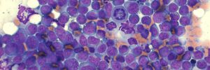
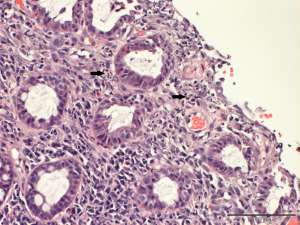
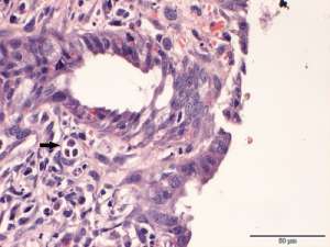
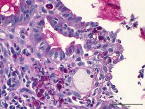
Moderate lymphoplasmacytic and eosinophilic colitis with mild to moderate, multifocal mucosal fibrosis and intra-mucosal algae
Discussion
Algal infection in dogs is an uncommon to rare occurrence and is often associated with Chlorella spp or Prototheca spp, a non-chloroplast containing variant of Chlorella. These organisms are ubiquitous in the environment and have been identified on all continents other than Antarctica. Attempts at recreating disease have met with varied success, suggesting that these are opportunistic pathogens that require some degree of immune suppression/tissue defect to enable infection. Most veterinary infections are cutaneous, reflecting secondary infection of open wounds or implantation of the organisms into the skin. In dogs, more disseminated infection can be seen and often involves the intestine and eyes. Ingestion of the organisms from the environment and presumed opportunistic infection of the intestinal tract (predominantly the colon) occurs. The organisms invade mucosal blood vessels and spread systemically. The observed retinal detachment in this dog was presumed to be related to the spread of infection to the eye. Definitive differentiation of these organisms is often difficult by histopathology alone. Histochemical or immunohistochemical stains, PCR, culture and/or electron microscopy can be used to help differentiate. The PAS stain used to highlight the organisms reveals smooth capsular staining without the presence of distinct starch granules. This would have me favour Prototheca infection in this dog.