
Osteoma cutis in a dog
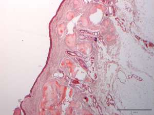
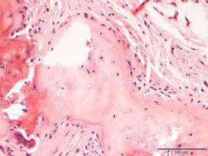
An 11-year-old dog presented to the referring vet with a history of multiple firm cutaneous plaques over the proximal limbs. The dog had been receiving long term prednisolone for the treatment of allergic skin disease. The referring vet submitted multiple skin biopsies for histopathology.
Final diagnosis
Osteoma cutis
Discussion
Osteoma cutis is defined by the formation of bone in the dermis or subcutis. Calcinosis cutis is a more common but related condition, that is associated with deposition of free mineral in the skin. Both osteoma and calcinosis cutis in the dog are most commonly associated with hyperglucocorticoidism, either iatrogenic or endogenous. In cases of calcinosis cutis there are often areas of bone formation, but there are also cases where bone formation is present in the absence of significant calcinosis, as was the case in this dog. In comparison to calcinosis cutis, cases of osteoma cutis may have much less inflammation, because bone, as opposed to free mineral, does not incite a significant tissue reaction.