Ocular manifestation of lymphoma in a cat
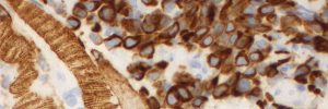
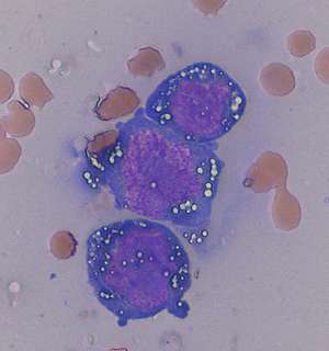
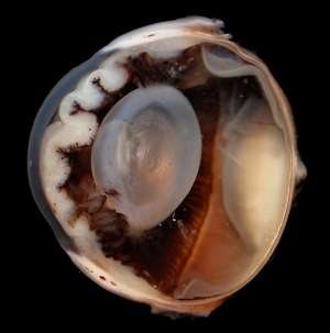
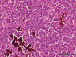
A 12-year-old male DSH cat presented with bilateral uveitis and diffuse thickening of the iris associated with a shallow anterior chamber. Below are images from the aqueocentesis sample (fig.1) and subsequent gross and histopathological specimens (figs.2&3) from one of the enucleated globes
Final Diagnosis
Uveal lymphoma (large cell, high grade)
Discussion
Cases of large cell/high grade lymphoma affecting the eye in both dogs and cats should be viewed as a multisystemic disease process even if the initial clinical manifestation appears to be restricted to the eye(s). Primary ocular lymphoma can occur but this is less common. Aqueocentesis can be used as a diagnostic tool in an attempt to confirm the diagnosis and is probably most useful when lymphoma is suspected. Enucleation can also be used to confirm the diagnosis in an irreversibly blind eye but should not be viewed as a curative procedure in the majority of cases.