Intraosseous epidermoid cyst in the toe of a dog
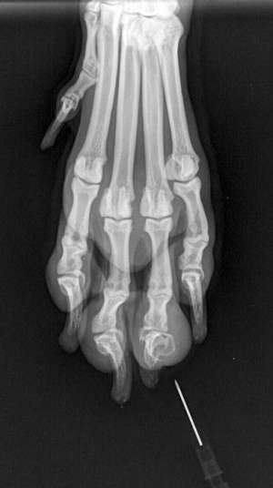
The fourth digit of the left foreleg was amputated in a 3-year-old, female Labrador Retriever in which radiographs indicated a focal expansile lytic lesion affecting the third phalanx (P3).
Final Diagnosis
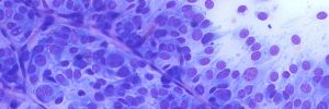
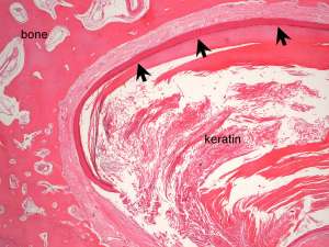
Intraosseous epidermoid cyst of the third phalanx
Discussion
Intraosseous epidermoid cysts are not common but account for lytic lesions occuring most commonly in the distal phalanges of dogs. Other sites occasionally affected include the vertebrae and skull. The cause of these benign cysts is not known but the main thoughts include nests of heterotopic ectoderm sequestered within bone during embryonic development or a penetrating wound resulting in traumatic implantation of epidermis into the underlying bone.