Intestinal pseudo-obstruction in a dog
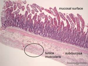
These are biopsy specimens taken at exploratory laparotomy from a thin-walled, distended small intestine in 6-year-old female Border Collie with ileus.
Final Diagnosis
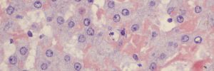
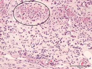
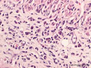
Lymphoplasmacytic intestinal leiomyositis associated with marked atrophy and loss of the tunica muscularis
Discussion
The histopathology in this case is consistent with so-called 'intestinal pseudo-obstruction', a rare disease that typically presents with ileus in the absence of mechanical/physical obstruction. Affected intestine is typically distended and thin-walled. It is possible that other organs containing smooth muscle may be affected (e.g. urinary bladder). The cause of the disease in dogs is not known but in human patients, the disease is thought to be due to a primary autoimmune disease process.