Hodgkin's-like lymphoma in a cat
A firm encapsulated, subcuticular mass was removed surgically in its entirety from the ventral neck of a 12-year-old domestic shorthair, male neutered cat.
Final Diagnosis
Lymphoma
Discussion
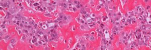
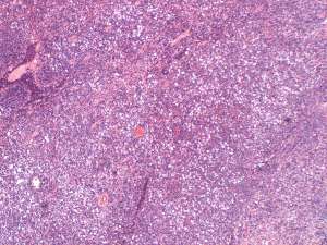
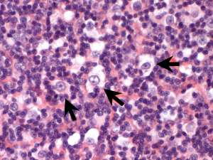
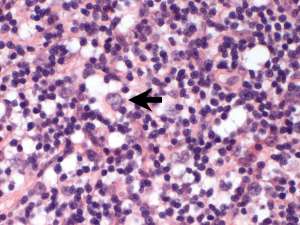
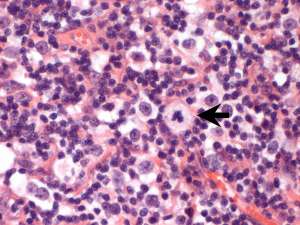
Histologically, this is consistent with a T cell-rich large B-cell lymphoma whereby the neoplastic cells make up a minor proportion of the lymphocyte population. In some cases, this can therefore make the diagnosis difficult on preliminary fine needle aspirates and small incisional/trucut biopsies. A typical presentation in cats is that of a single large lymph node in the cervical region (mandibular/neck location) that is firm but well-encapsulated and can therefore be removed in its entirety. Additional masses may occur in the same area as successive lymph nodes become involved over time. This bears some resemblance to Hodgkin's lymphoma in human patients and has acquired the name of so-called Hodgkin’s like lymphoma. This is a rare form of lymphoma in domestic animals and is typically associated with slow, low grade progression.