Fungal keratitis and secondary endophthalmitis in a horse
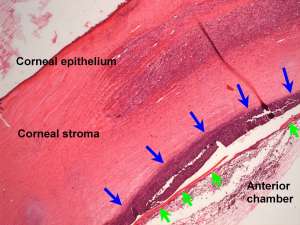
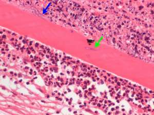
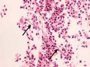
Enucleation of the right eye was performed in a 10-year-old male thoroughbred with persistent corneal ulceration, keratitis and uveitis. The following are H&E sections from the formalin-fixed globe.
Final Diagnosis
Fungal keratitis and endophthalmitis
Discussion
Fungi are opportunistic pathogens of the equine cornea and usually involve saprophytic fungi that are part of the resident fungal population found around the eye/adnexae. Species most commonly involved include Aspergillus spp, Alternaria spp. Penicillium spp. and Fusarium spp. Corneal injury is a common predisposing cause and apparent increased risk of fungal keratitis is also associated with increasing age, topical corticosteroid treatment and prolonged antibiotic therapy. Corneal abscesses secondary to fungal infection are often deep and therefore superficial corneal scrapes may yield false negative results. Perforation of Descemet’s membrane and secondary mycotic endophthalmitis such as what has occurred in this particular case, is a possible complication.