
Cutaneous cowpox virus infection in a cat
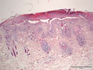
Multiple necro-ulcerative skin lesions on the forelimb in a 14-year-old DLH were excised and submitted for histopathology.
Final Diagnosis
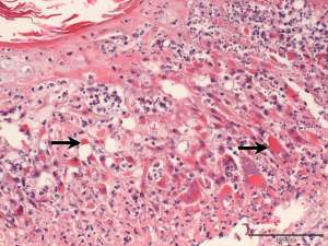
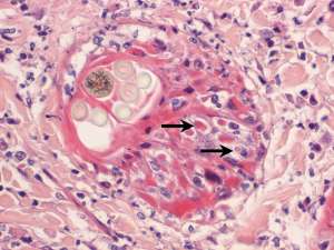
Necro-ulcerative dermatitis associated with abundant keratinocyte intracytoplasmic inclusion bodies typical of poxvirus infection
Discussion
Findings indicate cutaneous poxvirus infection, consistent with cowpox virus infection. The virus is typically carried by small mammals (voles, mice) and cats are usually infected via bite wounds. In most cases, lesions are self-limiting and are restricted to the skin and oral cavity, however systemic spread may occasionally occur (e.g. necrotising pneumonia). Unfortunately, lung infection with cowpox virus did occur in this cat (confirmed at post-mortem) and this is the result of systemic spread rather than primary respiratory infection. Cat to cat and zoonotic spread may occur however this is uncommon - young/old or immunocompromised individuals are at a higher risk of infection.