Bilateral ovotestes in a dog
A 2-year-old Cocker Spaniel presented for routine ovariohysterectomy. At surgery the uterus appeared small and contracted.
Final Diagnosis
Bilateral Ovotestes
Discussion
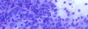
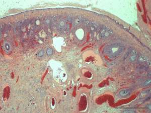
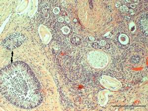
The presence of both male and female gonadal tissue in the ovaries of this dog is consistent with ovotestes. This is a feature that would indicate hermaphroditism. The exposure of hormones produced by the male gonadal tissue during embryogenesis (notable Mullarian Inhibitory Substance) will have resulted in hypoplasia/atrophy of the uterine tissue. The likely accounts for the reported small convoluted appearance of the uterus at the time of ovariohysterectomy. Ovariohysterectomy should be curative of any problems that may have arisen from this condition.