
Autumn has arrived - fatal cowpox infection in a cat
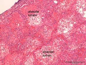
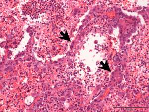
These are histopathology sections taken from a consolidated lung lobe in a 3-year-old male domestic shorthair cat that died following acute onset dyspnoea.

Final Diagnosis
Necrotising interstitial pneumonia associated with poxvirus infection
Discussion
The signalment and histopathological findings are consistent with cowpox infection. Despite its name, cowpox infection in cattle is relatively uncommon. The main reservoir hosts for the virus include small rodents and voles. Cats are typically infected following contact with the reservoir hosts and infection rates are often at their highest in Autumn when the concentration of rodents has reached its peak. The most common sign of cowpox infection in cats is a focal proliferative and necrotising dermatitis that frequently affects the face and extremities, correlating to the sites of bite wounds. Respiratory infection, presumed to arise secondary to systemic spread, is a less common manifestation and may be fatal in some cases. The lung pathology is often lobar in its distribution.